Patient’s details:
- Species: horse
- Sex: gelding
- Age: 11 years old
- Breed: Polish half bred horse
- Colour: bay
- Jumping obstacles
Medical history
Small oedema was visible half the length of the metacarpus of the left thoracic limb on the palmar side. The palpation showed an increased body surface temperature in the area of oedema. No lameness was found in the horse’s examination in motion. The ultrasound examination showed the injury to the tendon of the flexor digitorum superficialis in the metacarpal B region. Injury type: focused; injury class according to Genovese: 2, injury magnitude: 26%.
Treatment
The horse underwent a ten-day oral anti-inflammatory therapy. Hydrotherapy involving cold water massages (twice a day for 20 minutes) and kinesitherapy involving horse’s moving at a walk (twice a day for 20 minutes) were taken on. After the acute phase of the inflammation ended, two injections (at an interval of one month) of hyaluronic acid into the affected area (under the ultrasound control) were administered.
High power laser therapy
The high power laser therapy was given three months after the tendon injury. The procedure was carried out using the Polaris HP high power laser therapy equipment made by Polish company Astar.
Parameters of the procedure for wavelengths of 808 nm and 980 nm:
- Power (W): 4
- Frequenncy (Hz): 1000
- Radiation mode: Pulse
- Duty factor (%): 65
- Dose: (J/cm2): 15
The laser beam was applied across the entire tendon of the flexor digitorum superficialis running in the metacarpus of the left thoracic limb. Four therapeutic sessions at 24-hour intervals, three therapeutic sessions at 48-hour intervals and five therapeutic sessions at 96-hour intervals were performed. During the laser therapy, a trot was introduced into motor rehabilitation (saddled) on the hard ground. The trot duration was gradually increased from 2 to 5 minutes twice a day.
High power laser therapy: effects
During the high power laser therapy, three follow-up ultrasound examinations of the tendon healing process were carried out. Each time, the progress in the injury regeneration was observed. The injury was more hyperechoic; the longitudinal section showed forming bands of the collagen tissue. After the laser therapy ended, the injury was almost invisible in the ultrasound imaging.
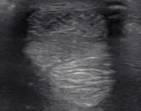
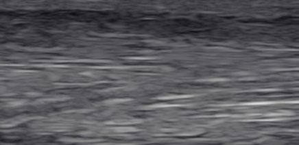
Fig. 1: Ultrasound image of the injury to the tendon of the flexor digitorum superficialis before the therapy.
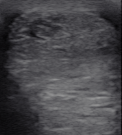

Fig. 2: Ultrasound image after the second injection of hyaluronic acid into the injury.
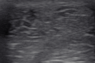
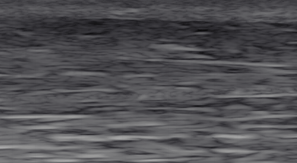
Fig. 3: High power laser therapy: ultrasound image before the first session.
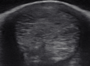
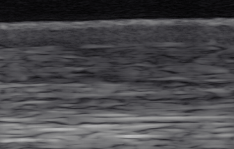
Fig. 4: High power laser therapy: ultrasound image after the 8th session.
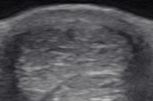
Fig. 5: High power laser therapy: ultrasound image after the sessions ended.
