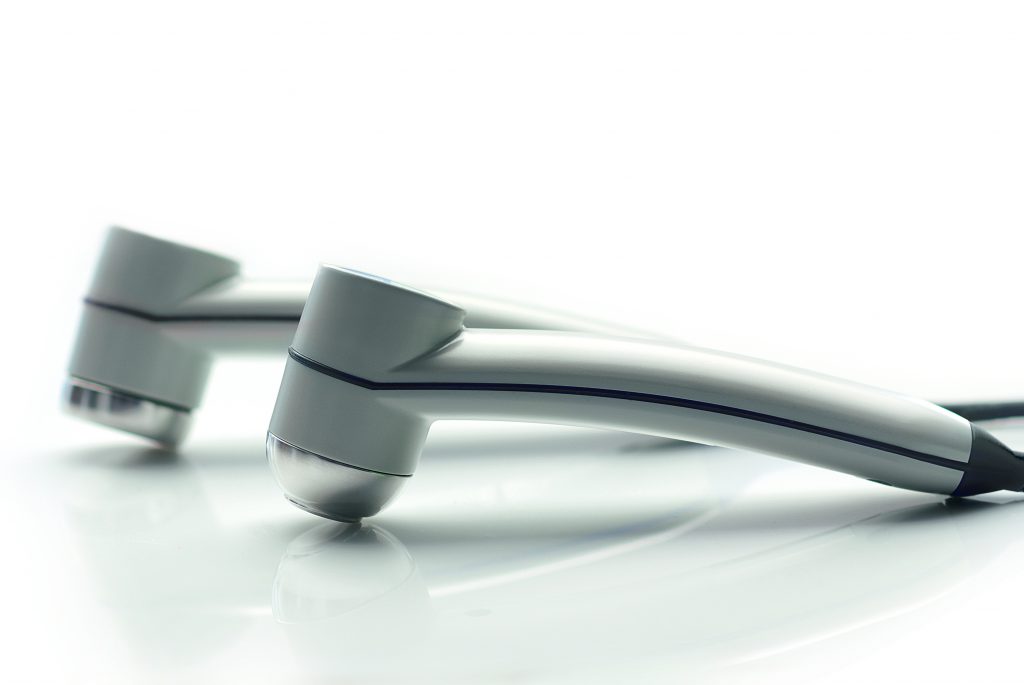
Ultrasounds are a frequently used treatment in physiotherapy. Ultrasound therapy is also known as sonotherapy. What does such a procedure look like and what equipment is used?
The article contains:
- Ultrasound therapy unit
- Methodology of performing the treatment with ultrasounds.
- Treatment parameters.
- How often should one perform an ultrasound treatment?
Ultrasounds – portable ultrasound therapy unit
The Astar company has prepared a range of several units for ultrasound therapy:
- PhysioGo.Lite Sono
- PhysioGo.Lite Combo
- PhysioGo 200A
- PhysioGo 300A
An example unit for ultrasound treatments is ASTAR’s PhysioGo 200A. The unit allows ultrasound therapy and phonophoresis to be performed in continuous or pulsed mode.
The PhysioGo 200A works with 1 MHz or 3.5 MHz ultrasound heads with an effective radiation area of 1 cm2 or 4 cm2.
Discover what the treatment looks like when using the portable ultrasound therapy device, the PhysioGo 200A.
Methodology of performing the treatment with ultrasounds
How to perform an ultrasound therapy treatment? For the treatment to be effective, all parameters need to be adapted individually to the patient’s condition. The treatment methodology can be divided into several stages.
Ultrasound therapy – principles of application:
– Choosing the coupling agent,
– Choosing the head guidance method,
– Choosing the application method,
– Adjusting the parameters

Choosing the coupling agent
In order to perform the treatment properly, a suitable ultrasound conductive agent should be used, e.g. gel. It is applied directly to the surface of the skin.
Choosing the head guidance method
The treatment is performed using the ultrasound head Depending on the type of surface to be treated, the method of moving the head is chosen:
- labile technique, during which the physiotherapist moves the head continuously, parallel to the skin. Movements are performed at an average speed of 4cm/s in the following way:
– with overlapping circles – for irregularly shaped surfaces;
– with longitudinal movements – for larger and flat surfaces.
- the water immersion method, which is used when the treated area (e.g. foot) is irregularly shaped.
Choosing the application method
The physiotherapist has a choice of application methods such as:
- direct (local) method – treatment performed in direct contact with the tissue,
- indirect (segmental) method – refers to the treatment performed in the spinal region.
Treatment parameters
Some of the parameters that influence the selection of the appropriate ultrasound dose are:
Mode of operation
- continuous
- pulse
Frequency
- 1 MHz – used for the treatment of musculo-fascial trigger points and those located in connective tissue,
- 3.5 MHz – used for surface point therapy of the skin.
Intensity
- 5 W/cm2 – used during severe pain, in the face and neck area,
- 5 – 1.0 W/cm2 – used for the treatment of medium intensity low back area pain,
- 0 – 1.5 W/cm2 – recommended for low intensity pain in the hip, buttock and limb areas.
Duty factor
The most common is 20 -75% infill.
- How often to use ultrasound – how long does the treatment last?
Depending on the type of condition being treated or the size of the area to be hyperacoustic, the duration of the treatment, the number of treatments in a series and the frequency varies.
Treatment time
- short: 1-3 min
- medium: 4-9 min
- long: 10-15 min
- in the segmental method: the time should not exceed 2 min.
Number of treatments in a series
The number of treatments set by the physiotherapist is between 3 and 15 in a series.
Frequency of treatments
Typically, in acute conditions, treatments are performed daily and in chronic conditions every two days.
See examples of parameters and frequency of treatments – ankle sprain
Ankle sprain – subacute condition |
||
| Carrier frequency | 1 MHz | |
| Impulse frequency | 100 Hz | |
| Duty factor | 50% | |
| Power density | 1.2 W/cm² | |
| Ultrasound head | 4 cm2 | |
| Treatment time | 8 min | |
| Number of treatments in a series | 5 – 10 | |
| Frequency of treatments: | Daily | |
| Research methods: | Labile technique, sonication area – locally around the joint | |
| Effect | Increased tensile strength of collagen fibres, reduced tension, increased temperature in tissues, hyperaemia, pain relief | |
